Skin Surgeons Edinburgh 0131 376 2744
SKIN SURGEONS Make an enquiry
Patient guide · Edinburgh

Understanding Basal Cell Carcinoma

Patient guide · Basal cell carcinoma

An educational guide to basal cell carcinoma.

Basal cell carcinoma (BCC) is the most common type of skin cancer in the UK, usually caused by long-term exposure to ultraviolet (UV) radiation from the sun or tanning beds. BCC rarely spreads to other parts of the body, but it can damage surrounding tissue if left untreated, so early diagnosis and professional treatment matter. This guide explains the warning signs, causes, types, treatment options, and prevention.

Concerned about a lesion? Visit our skin cancer service in Edinburgh

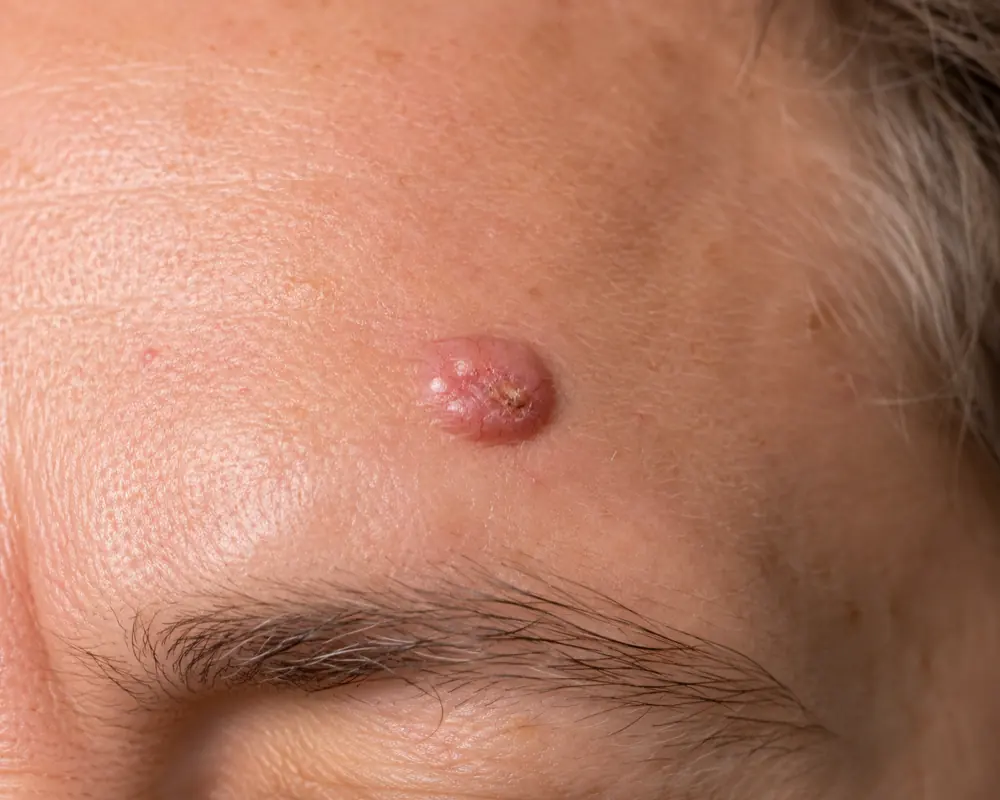
A basal cell carcinoma lesion on the skin of the face
The basics

What is basal cell carcinoma?

Basal cell carcinoma is a slow-growing form of skin cancer that arises from the basal cells in the epidermis. These cells normally produce new skin cells, but when their DNA is damaged they can grow uncontrollably and form a tumour.

  • It is the most common type of skin cancer in the UK.
  • BCC typically develops on sun-exposed areas such as the face, neck, and hands.
  • Although BCC rarely spreads, it can grow deeper into the skin and cause disfigurement if left untreated.

Read more on basal cell carcinoma at the British Association of Dermatologists.

When to seek help

Warning signs of basal cell carcinoma

BCC can present in several ways, so it's important to recognise the early signs:

  • Shiny bump or nodule: often pearly or translucent, with visible blood vessels.
  • Red or scaly patch: may look irritated or resemble eczema.
  • Non-healing sore: a wound that doesn't heal and may bleed or crust over.
  • Scar-like lesion: a flat, flesh-coloured or white area resembling a scar, often with poorly defined edges.

If you notice any persistent change to your skin — a lesion that grows, bleeds, or fails to heal — have it assessed by a specialist promptly.

Causes

Causes and risk factors

The primary cause of BCC is UV radiation, which damages the DNA of skin cells and leads to abnormal growth. Common risk factors include:

  • Sun exposure: long-term exposure to the sun, or use of tanning beds.
  • Fair skin: people with light skin, hair, and eyes, or who burn easily, are at greater risk.
  • Age: BCC is more common over 50, although younger people can be affected.
  • Previous skin cancer: a history of BCC increases the chance of another lesion.
  • Weakened immune system: conditions or medications that suppress immunity increase susceptibility.
Why treat

Why treat basal cell carcinoma?

While BCC is not usually life-threatening, treatment is important to prevent complications:

  • Local tissue damage: untreated BCC can invade deeper tissue, including muscle and bone.
  • Disfigurement: lesions on the face or other visible areas can cause scarring and cosmetic concern.
  • Recurrent lesions: treating BCC early reduces the risk of recurrence or a larger, harder-to-treat tumour.
Types

Types of basal cell carcinoma

There are several types of BCC, each with its own characteristics:

  • Nodular BCC: a shiny, pearl-like bump — the most common presentation.
  • Superficial BCC: a red, scaly patch often found on the trunk or limbs.
  • Morphoeic (morpheaform) BCC: an aggressive type that resembles a scar and can infiltrate deeper tissue.
  • Pigmented BCC: contains dark pigment and may resemble a melanoma.

Because a pigmented BCC can look like a melanoma, any dark or changing lesion should be assessed — see our melanoma guide for the warning signs.

Treatment options

Treatment options explained

Treatment for BCC depends on the size, type, location, and depth of the tumour. The main options are:

  • Surgical excision: the lesion is removed with a margin of healthy skin. High cure rate and precise — the usual treatment for most BCCs.
  • Topical medications: prescription creams or gels. Non-invasive — mainly for selected superficial, low-risk BCCs, or patients who cannot have surgery.
  • Cryotherapy: the lesion is frozen off. Quick and minimally invasive — mainly for selected small, superficial, low-risk BCCs.
  • Mohs micrographic surgery: a specialist technique that removes the tumour in stages with the margins checked under the microscope as it proceeds. It may be appropriate for selected high-risk, recurrent, or difficult-site BCCs — particularly on the face — and is usually arranged through a specialist Mohs service.
  • Radiotherapy: targeted radiation. Used in selected cases, often for patients unable to undergo surgery.

Excised tissue is sent for histological (laboratory) analysis as standard, to confirm the diagnosis and check the margins are clear.

What you pay depends on the diagnosis and the complexity of the lesion — a specialist consultation is £200, and removal pricing is confirmed once the diagnosis is known. See full pricing and arrange a skin cancer assessment in Edinburgh.

During & after

What to expect during treatment

At your consultation, a skin cancer specialist examines the lesion and may take a biopsy to confirm the diagnosis if there's any doubt. A tailored treatment plan is then discussed based on the type and size of the BCC, the procedure is carried out, and regular follow-up checks confirm the lesion has been treated and monitor for recurrence.

Aftercare tips

  • Keep the area clean: wash gently with mild soap and water once your dressings are removed.
  • Protect from sunlight: use sunscreen and avoid sun exposure to prevent further damage.
  • Follow specialist advice: use any prescribed ointments or dressings as directed.

For full preparation and recovery guidance, read our what to expect page.

Prevention

Preventing basal cell carcinoma

You can reduce your risk of developing BCC by:

  • Wearing broad-spectrum sunscreen daily (SPF 30 or higher).
  • Avoiding peak sun hours (10am to 4pm).
  • Wearing protective clothing, hats, and sunglasses.
  • Avoiding tanning beds.
  • Performing regular self-checks for changes to your skin.
Expertise

Why choose a plastic surgeon for BCC removal

See a specialist if you notice a persistent sore that doesn't heal, a change in the size, shape, or colour of a lesion, or if you have a history of prolonged sun exposure or previous skin cancer.

Plastic surgeons are uniquely equipped to deliver the best cosmetic and cancer-safe outcome. With advanced training in skin surgery and aesthetics, they can minimise scarring while ensuring the lesion is fully removed. At Skin Surgeons Edinburgh, every procedure is carried out by Dr Kazem Nassar, a Consultant Plastic Surgeon on the GMC Specialist Register, at the Waterfront Private Hospital.

Dr Kazem Nassar is the plastic surgery lead for the South East Scotland melanoma multidisciplinary team (MDT) and also sits on the South East Scotland non-melanoma skin cancer MDT, providing specialist surgical input into the assessment and management of melanoma, non-melanoma skin cancer and complex skin cancer cases.

Frequently asked

Skin cancer FAQs

Skin cancer FAQ

01 What is skin cancer surgery?

Skin cancer surgery removes cancerous lesions — including basal cell carcinoma (BCC), squamous cell carcinoma (SCC), and melanoma. The aim is complete removal while preserving as much healthy tissue as possible.

02 What treatment techniques are used for skin cancer?

Several techniques are used, surgical and non-surgical. Surgical excision removes the cancerous tissue with a margin of healthy skin and is the usual treatment for most invasive skin cancers. Curettage and cautery, topical prescription creams, or cryotherapy may be used for selected low-risk or superficial lesions. The right choice depends on the type of cancer, its risk features, and where it is — your surgeon will explain the options.

03 What are the potential risks of skin cancer treatment?

As with any procedure there are risks. These can include bleeding, infection, problems with wound healing, scarring, altered sensation around the wound, incomplete excision (cancer cells left at the edge of what was removed), and recurrence — any of which may mean further treatment is needed. Your surgeon discusses the risks relevant to your lesion and takes precautions to reduce them.

04 Will there be scarring after treatment?

Scarring depends on the size and location of the lesion and the treatment method used. The surgeon aims for the best possible cosmetic outcome, particularly in cosmetically sensitive areas such as the face.

05 How long does skin cancer treatment take?

The procedure typically lasts from about 30 minutes to a few hours, depending on the complexity and type of treatment.

06 Is skin cancer treatment painful?

Local anaesthetic is applied to the area to prevent pain during surgical treatment. You may feel minor discomfort or a sensation of pressure during and after the procedure.

07 Can I resume normal activities after the procedure?

Most people return to their daily activities shortly afterwards, though recovery time varies — especially for larger or more complex surgery.

08 When should I consider skin cancer treatment?

Seek assessment if you've been diagnosed with BCC, SCC, or melanoma, or if you have a suspicious lesion that is changing in size, shape, or colour, or that bleeds, itches, or causes pain. A changing or non-healing lesion should be seen promptly.

09 How is the skin cancer analysed after removal?

Removed tissue is sent for histological (laboratory) analysis as standard, to confirm the diagnosis and check that all cancerous cells have been removed. This guides whether any further treatment is needed.

10 How do I choose between different treatment options?

The choice depends on the type, size, and location of the cancer, along with your medical history and cosmetic concerns. Your surgeon will discuss the most appropriate option at your consultation.

11 What should I expect in terms of aftercare and healing?

Aftercare involves keeping the treatment site clean, applying any prescribed ointment, and following your surgeon's instructions. Healing time varies but usually takes a few weeks.

12 Can skin cancer recur after treatment, and is follow-up needed?

Although the goal is to remove all cancerous cells, there is a chance of recurrence. A follow-up appointment is usually arranged to check healing and review the histology results. Ongoing surveillance then depends on the diagnosis, the risk features of the lesion, any previous skin cancer history, and MDT advice.

Ready when you are

Arrange a skin cancer assessment.

A changing or non-healing lesion should be seen promptly. Our consultant-led service provides specialist assessment, diagnosis, and treatment, with histology as standard. No GP referral required.

Prefer to talk? Call 0131 376 2744.