Skin Surgeons Edinburgh 0131 376 2744
SKIN SURGEONS Make an enquiry
Patient guide · Edinburgh

Understanding Melanoma

Patient guide · Melanoma

An educational guide to melanoma.

Melanoma is one of the most serious forms of skin cancer. It arises from melanocytes, the cells that produce pigment in the skin. Although it accounts for a smaller share of skin cancers than BCC or SCC, it carries a greater risk of spreading and can be life-threatening if left untreated — so early diagnosis and treatment really matter. This guide covers the ABCDE warning signs, causes, how melanoma is diagnosed, treatment options, and prevention.

Concerned about a mole or lesion? Visit our skin cancer service in Edinburgh

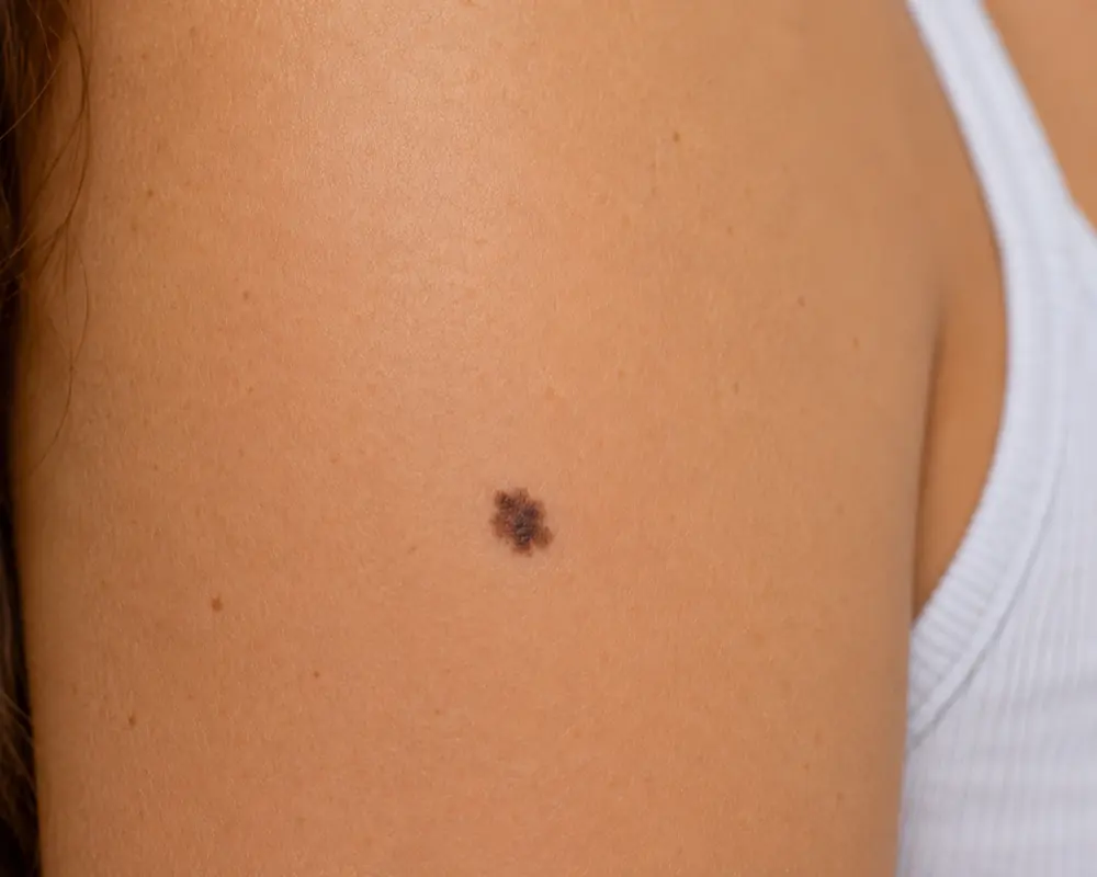
A melanoma skin lesion showing irregular borders and colour
The basics

What is melanoma?

Melanoma develops when melanocytes are damaged — typically by ultraviolet (UV) radiation — and begin to grow uncontrollably. It can appear anywhere on the body, including areas not exposed to sunlight such as the soles of the feet, the palms, or under the nails.

Types of melanoma

  • Superficial spreading melanoma: the most common type, often starting as an irregular, flat mole that spreads outwards before growing deeper.
  • Nodular melanoma: a more aggressive type that appears as a raised, dark lesion and grows rapidly.
  • Lentigo maligna melanoma: usually seen in older people, developing slowly on sun-exposed areas such as the face.
  • Acral lentiginous melanoma: a rarer type, more common in darker skin tones, appearing on the hands, feet, or under the nails.

Read more on melanoma at Melanoma Focus.

When to seek help

ABCDE warning signs of melanoma

Recognising melanoma early is critical. Use the ABCDE rule to check a mole or skin change:

  • A — Asymmetry: one half of the mole doesn't match the other.
  • B — Border: irregular, scalloped, or poorly defined edges.
  • C — Colour: uneven colour, including shades of brown, black, red, or white.
  • D — Diameter: larger than 6 mm, although smaller melanomas can still be dangerous.
  • E — Evolving: any change in size, shape, or colour, or symptoms such as itching or bleeding.

Melanoma may also appear as a new, unusual growth or a change in an existing mole. If you notice any of these signs, seek medical advice without delay. Our mole guide explains the ABCDE check in more detail.

Causes

Causes and risk factors

Melanoma is primarily caused by UV radiation, from sunlight or artificial sources such as tanning beds, but genetic and lifestyle factors also contribute. Key risk factors include:

  • Sunburns: a history of severe sunburn, particularly in childhood.
  • Tanning beds: regular use significantly raises melanoma risk.
  • Fair skin: fair skin, freckles, and light hair carry greater risk.
  • Family history: a close relative with melanoma raises your risk.
  • Multiple moles: many moles, or atypical (dysplastic) moles, are a significant risk factor.
  • Immune suppression: conditions or treatments that weaken the immune system increase susceptibility.
Diagnosis

How is melanoma diagnosed?

Diagnosis usually begins with a thorough skin examination by a specialist. If melanoma is suspected:

  • Dermoscopic examination: a magnified view of the lesion helps identify suspicious features.
  • Excision biopsy: the whole lesion is removed with a narrow margin and sent for histological (laboratory) analysis to confirm the diagnosis and measure key features such as depth.
  • Imaging tests: in higher-risk or advanced cases, imaging may be used to check whether melanoma has spread to lymph nodes or elsewhere.

If melanoma is confirmed, the histology guides the next steps — which may include a wider excision, a sentinel lymph node biopsy, imaging, oncology treatment, or transfer into the NHS melanoma pathway for staging and ongoing management.

Treatment options

Melanoma treatment options explained

The primary treatment for melanoma is surgical removal; the exact approach depends on the stage and location.

Surgical techniques

  • Excision biopsy: the whole suspected melanoma and a narrow margin are removed first, to confirm the diagnosis on histology — the usual first step.
  • Wide local excision: once melanoma is confirmed, more surrounding tissue is removed; the margin depends on the depth of the melanoma.
  • Sentinel lymph node biopsy: a staging and prognostic test offered for selected higher-risk melanomas to check whether nearby lymph nodes are involved — it is not the main treatment itself.

Advanced treatments

  • Immunotherapy: medications that boost the immune system to attack melanoma cells.
  • Targeted therapy: for melanomas with specific genetic mutations, targeting abnormal proteins in cancer cells.
  • Radiotherapy: used for advanced melanomas or where surgery is not feasible.

These later steps — wider excision, sentinel lymph node biopsy, imaging, and oncology treatment — are generally delivered through the NHS melanoma pathway (see below).

Excised tissue is sent for histological (laboratory) analysis as standard. What you pay depends on the diagnosis and complexity — a specialist consultation is £200, and the cost of removing a suspicious lesion is confirmed once it has been assessed. See full pricing and arrange a skin cancer assessment in Edinburgh.

During & after

What to expect during initial treatment

At your consultation, a skin cancer specialist examines the lesion and may take a biopsy to confirm the diagnosis. Based on the result, a tailored plan is discussed and an excision biopsy is performed. Regular follow-up checks confirm the lesion has been treated and monitor for recurrence; further tests may be needed depending on the biopsy result.

Your care may be discussed at the local NHS melanoma multidisciplinary meeting, and may be transferred into the NHS if further surgery or staging tests are required. This shared pathway makes sure a confirmed melanoma is managed by the full specialist cancer team.

Aftercare tips for skin biopsies

  • Keep the area clean: wash gently with mild soap and water once your dressings are removed.
  • Protect from sunlight: use sunscreen and avoid sun exposure to prevent further damage.
  • Follow specialist advice: use any prescribed ointments or dressings as directed.

For full preparation and recovery guidance, read our what to expect page.

Prevention

Preventing melanoma

You can reduce your risk of melanoma — and catch it early — with a few habits:

  • Use sunscreen daily: broad-spectrum, SPF 30 or higher, reapplied every two hours and after swimming or sweating.
  • Limit UV exposure: avoid direct sun at peak hours (10am to 4pm), wear a wide-brimmed hat, long sleeves, and UV-blocking sunglasses, and avoid tanning beds.
  • Check your skin monthly: head to toe, including the scalp, soles, and under the nails, using the ABCDE signs above.
  • Have professional skin checks: a full-body skin exam, with the frequency your clinician advises if you have risk factors such as fair skin or a family history.
  • Protect children: sunscreen, protective clothing, and shade reduce lifetime UV exposure.
  • Stay vigilant: be especially watchful if you've had melanoma or another skin cancer before.
Expertise

Why choose a plastic surgeon for melanoma removal

See a skin specialist if you notice a change in a mole or lesion, a new or unusual skin growth, or any lesion that starts to itch, bleed, or cause pain — seek advice promptly. Early consultation leads to timely diagnosis and treatment, which improves outcomes.

Choosing a plastic surgeon for melanoma removal means the procedure is carried out with precision, prioritising both complete cancer removal and a good cosmetic and functional result. Plastic surgeons are highly trained in skin cancer excision and reconstructive techniques. At Skin Surgeons Edinburgh, every procedure is carried out by Dr Kazem Nassar, a Consultant Plastic Surgeon on the GMC Specialist Register, at the Waterfront Private Hospital.

Dr Kazem Nassar is the plastic surgery lead for the South East Scotland melanoma multidisciplinary team (MDT) and also sits on the South East Scotland non-melanoma skin cancer MDT, providing specialist surgical input into the assessment and management of melanoma, non-melanoma skin cancer and complex skin cancer cases.

Frequently asked

Skin cancer FAQs

Skin cancer FAQ

01 What is skin cancer surgery?

Skin cancer surgery removes cancerous lesions — including basal cell carcinoma (BCC), squamous cell carcinoma (SCC), and melanoma. The aim is complete removal while preserving as much healthy tissue as possible.

02 What treatment techniques are used for skin cancer?

Several techniques are used, surgical and non-surgical. Surgical excision removes the cancerous tissue with a margin of healthy skin and is the usual treatment for most invasive skin cancers. Curettage and cautery, topical prescription creams, or cryotherapy may be used for selected low-risk or superficial lesions. The right choice depends on the type of cancer, its risk features, and where it is — your surgeon will explain the options.

03 What are the potential risks of skin cancer treatment?

As with any procedure there are risks. These can include bleeding, infection, problems with wound healing, scarring, altered sensation around the wound, incomplete excision (cancer cells left at the edge of what was removed), and recurrence — any of which may mean further treatment is needed. Your surgeon discusses the risks relevant to your lesion and takes precautions to reduce them.

04 Will there be scarring after treatment?

Scarring depends on the size and location of the lesion and the treatment method used. The surgeon aims for the best possible cosmetic outcome, particularly in cosmetically sensitive areas such as the face.

05 How long does skin cancer treatment take?

The procedure typically lasts from about 30 minutes to a few hours, depending on the complexity and type of treatment.

06 Is skin cancer treatment painful?

Local anaesthetic is applied to the area to prevent pain during surgical treatment. You may feel minor discomfort or a sensation of pressure during and after the procedure.

07 Can I resume normal activities after the procedure?

Most people return to their daily activities shortly afterwards, though recovery time varies — especially for larger or more complex surgery.

08 When should I consider skin cancer treatment?

Seek assessment if you've been diagnosed with BCC, SCC, or melanoma, or if you have a suspicious lesion that is changing in size, shape, or colour, or that bleeds, itches, or causes pain. A changing or new pigmented lesion should be seen promptly.

09 How is the skin cancer analysed after removal?

Removed tissue is sent for histological (laboratory) analysis as standard, to confirm the diagnosis and check that all cancerous cells have been removed. This guides whether any further treatment is needed.

10 How do I choose between different treatment options?

The choice depends on the type, size, and location of the cancer, along with your medical history and cosmetic concerns. Your surgeon will discuss the most appropriate option at your consultation.

11 What should I expect in terms of aftercare and healing?

Aftercare involves keeping the treatment site clean, applying any prescribed ointment, and following your surgeon's instructions. Healing time varies but usually takes a few weeks.

12 Can skin cancer recur after treatment, and is follow-up needed?

Although the goal is to remove all cancerous cells, there is a chance of recurrence. A follow-up appointment is usually arranged to check healing and review the histology results. Ongoing surveillance then depends on the diagnosis, the risk features of the lesion, any previous skin cancer history, and MDT advice.

Ready when you are

Arrange a skin cancer assessment.

A new or changing pigmented lesion should be seen promptly. Our consultant-led service provides specialist assessment, biopsy, and diagnosis, with histology as standard and a clear onward pathway if a melanoma is confirmed. No GP referral required.

Prefer to talk? Call 0131 376 2744.